

[ad_1]

Scientists from Trinity College Dublin and the Royal College of Surgeons in Ireland have developed innovative color-changing fluorescent dyes to visualize different biological environments using a single dye. These dyes, capable of being “turned on” and “off” depending on their location within cellular structures, allow for real-time, high-contrast images of cellular processes. This breakthrough, published in the journal Chem, paves the way for advances in biosensing, drug delivery imaging, and the study of cell dynamics. The research benefits from international collaboration and significant funding from Irish research bodies, promising a wide range of applications in biology and medicine. Credit: SciTechDaily.com
Researchers at Trinity College Dublin, in collaboration with the Royal College of Surgeons of Ireland (RCSI), have developed special color-changing fluorescent dyes that, for the first time, can be used to simultaneously visualize multiple different biological environments using a single dye.
When these dyes are encapsulated in delivery containers, such as those used in technologies such as COVID-19 The vaccines “turn on” and emit light through a process called “aggregation-induced emission” (AIE). Shortly after their introduction into cells, their light is “turned off” before being “turned on” again once the cells transfer the dyes to cellular lipid droplets.
Advanced imaging techniques
Because light coming from inside cells is a different color and occurs within a different time window than light coming from the same dye inside blood vessels, researchers can use a technique called “life-long imaging.” fluorescence” (FLIM) to distinguish between the two environments in real time.
The work was recently published in the main international journal, Chemistry. First author Dr Adam Henwood, principal investigator from the School of Chemistry and Trinity Biomedical Sciences Institute (TBSI), worked on this design with PhD student Connie Sigurvinsson.
Dr Henwood explained: “Bioimaging is based on “on/off” dyes where the dyes only emit light under one set of conditions but otherwise turn off. This is extremely useful, but it means you can only look at one place at a time under the microscope. What is interesting about this work is that our dyes reach an optimal point that gives them distinctive on/off properties and, more importantly, we can observe and differentiate these different on states.
“So, we both see more and better than before. We do this by timing the time it takes for light from our samples to reach the microscope: the light from the delivery vessels takes a little longer than the light from inside the cells. By collecting enough light signals, we can use this information to quickly create accurate 3D images of the two different dye environments. “The time differences are small (just a few billionths of a second either way), but our method is sensitive enough to capture them.”
This unique quality means that the dyes could have a huge set of applications and, for example, have the potential to revolutionize biosensing and imaging approaches.
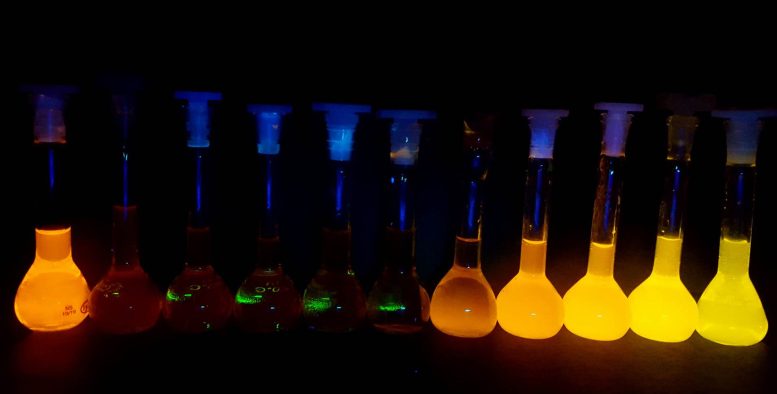
Luminescence changes of the same dye going from the pure organic solvent, left, to water, right. Credit: Dr Adam Henwood, Trinity College Dublin
Because these dyes can help scientists map the intricate structures within living cells with high contrast and specificity, they could help illuminate how cells absorb and metabolize drugs or allow scientists to design and perform a variety of new experiments to improve our understanding of the complex inner workings of cells and their all-important biochemical machinery.
In the paper published in the journal, the scientists focused on using dyes to image cellular lipid (fat) droplets, which are an example of important “organelles” that make up living cells in most complex organisms. (like us humans).
Lipid droplets, once considered simple “fat stores,” are now believed to play an important role in regulating cellular metabolism, coordinating the absorption, distribution, storage and use of lipids in cells. Because of this growing understanding of their importance, and because sudden changes in their activity often indicate cellular stress, they serve as a useful testing scenario for dyes. A possible avenue of additional research is to see if the team can target other important cellular organelles with their dyes.
Thorfinnur Gunnlaugsson, professor of chemistry in Trinity’s School of Chemistry and based at TBSI, is the lead author of the paper. He said:
“Being able to monitor cellular function or the flow of molecules or drug candidates within cells by observing different colors of fluorescence emission is extremely attractive. The big breakthrough here is that we can resolve and use the difference in their fluorescence lifetimes to identify these same probes within different cellular environments in a rapid and accurate manner, allowing us to literally chart their colorful “time travel” within the cells. cells.
“The most interesting thing, however, is that this phenomenon does not apply to cellular images. These results open new possibilities in everything from the study of chemical biology, as we have shown here, to many other medical applications and even in the generation of new functional materials for use beyond biology. “Any molecular or nanomaterial that requires controlled molecular motion can, in principle, be mapped and tuned using our new method.”
Potential applications and future directions
And, in fact, this is where the authors intend to cast the net far and wide. They envision many new possibilities for these dyes, noting that their exceptional sensitivity is attractive for developing sensors for hazardous environmental pollutants or using their bright, light-emitting properties to drive chemical transformations, analogous to those in nature. photosynthesis.
The research is both international (eight countries are represented) and Irish in character, with the latter’s main funding bodies, the Irish Research Council (IRC) and the Irish Science Foundation, playing key financial supporting roles. Most notable is the SFI Pharmaceuticals Research Centre, SSPC, which primarily funded the work, as well as contributions from the SFI AMBER center and through the EPSRC-SFI Center for Doctoral Training Program based at AMBER.
Prof Damien Thompson, Professor of Physics at the University of Limerick and Director of the SSPC, said: “As a centre, we continue to advance and create new knowledge at the interface of materials and biology. This collaborative work between two of our senior researchers at Trinity and RCSI shows the power of fundamental science to drive innovation in medicine. The closer we look at the molecule-cell interface and, more importantly, the better we can see, in real time, how molecules diffuse from place to place within the cellular nanomachinery, the closer we will be to making the Richard Feynman’s dream of understanding everything that living beings do from the movement and motion of atoms.
“But only recently have researchers had sufficient experimental and computational resources to track these movements and vibrations in complex biological environments. “This exciting new work demonstrates more specific, high-contrast imaging of subcellular dynamics, which in turn will allow researchers to develop more effective drug formulations with reduced side effects.”
Professor Donal O’Shea, who supervised the research, is a cellular imaging expert working in the Department of Chemistry and the RCSI Super Resolution Imaging Consortium (funded by Science Foundation Ireland, SFI). He added: “Our use of FLIM to track dynamic interactions of AIE with living cells is an approach that may have broad applicability to other fluorophore systems, allowing insights to be gained that were previously hidden.”
Reference: “Time-resolved fluorescence imaging with color-changing AIE “on/on” nanoparticles” by Adam F. Henwood, Niamh Curtin, Sandra Estalayo-Adrián, Aramballi J. Savyasachi, Tómas A. Gudmundsson, June I. Lovitt, L. Constance Sigurvinsson, Hannah L. Dalton, Chris S. Hawes, Denis Jacquemin, Donal F. O’Shea and Thorfinnur Gunnlaugsson, December 1, 2023. chemistry.
DOI: 10.1016/j.chempr.2023.10.001
The study was funded by the Irish Research Council and the Science Foundation of Ireland.